
Read full research article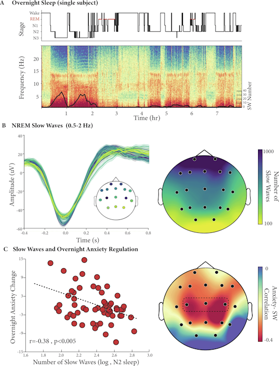
OVERVIEW
Aging takes a toll on more than just memory — it quietly erodes emotional stability. The most common mental health condition in adults over 60, anxiety doesn't just cause distress on its own: each new anxiety symptom nearly doubles the risk of progressing from mild cognitive impairment to Alzheimer's disease. Two brain changes reliably accompany aging — shrinkage in emotion-regulating brain regions and a steep decline in deep, slow-wave sleep — but until now, no one has examined whether those two factors are connected to ask whether the loss of deep sleep's characteristic "slow waves" is the missing link between an aging brain and worsening emotional health.Researchers recruited 61 cognitively healthy adults over age 65, each with varying levels of anxiety, and brought them into the sleep lab for a carefully monitored overnight stay. Electrodes recorded brain activity throughout the night, capturing the slow electrical oscillations that define deep, restorative sleep. The following morning, participants completed anxiety questionnaires and underwent MRI brain scans so researchers could measure tissue loss in regions known to govern fear and emotion — including the amygdala, insula and cingulate cortex. To check whether findings held up over time, a subset of 24 participants returned an average of four years later to repeat the entire process.The results tell a coherent and clinically important story. Participants who generated fewer slow waves during the night woke up more anxious the next morning — and this effect held steady even after accounting for age, sex and baseline anxiety tendencies. Brain scans confirmed older adults with more tissue loss in emotion-sensitive regions also produced fewer slow waves. Findings open a genuinely actionable door for future research: non-invasive techniques that boost slow-wave activity during sleep — such as acoustic or gentle electrical brain stimulation — may offer a way to preserve emotional resilience in aging brains, even in the presence of structural decline that cannot be reversed.Fig. 1: NREM Slow Waves and Anxiety.A Hypnogram of a single subject (top) with its corresponding multitaper spectrogram (Bottom, C3 channel). The superimposed black line denotes the number of detected slow wave events across the night (5 min averages). B Detected slow waves from NREM sleep (left panel, single subject example, minimum 100 detections per plotted channel) and average slow wave counts across participants (N = 61, right panel). C Increased number of slow waves during NREM2 sleep is associated with lower next-day anxiety (N = 58, R = -0.38, P < 0.005, central derivative, left panel) also evident across the scalp (right panel, predefined central derivative marked in a dotted line, only channels with a minimum of 20 detections are plotted).