Facebook pixel
Go to home page

State-of-the-Art Hyperscanning Technology

One of two Siemens 3T Prisma scanners utilizing Syngo MR E11 software at the Sammons BrainHealth Imaging Center, located inside Center for BrainHealth, part of UT Dallas.

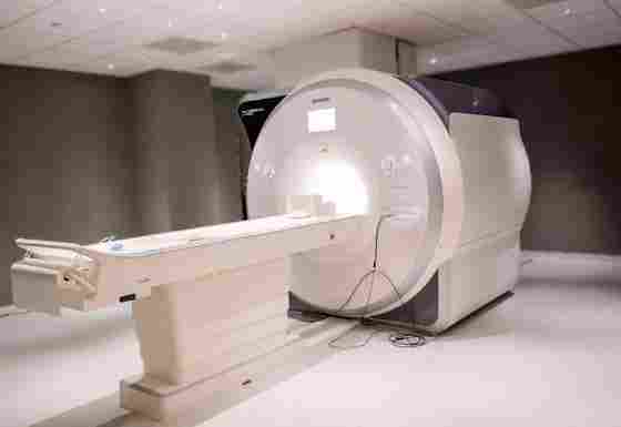
Each scanner has a 60-cm bore and is capable of accommodating up to 204 connected coil elements and 64 RF channels, with three coil configurations available for imaging: a 20-channel (16 head, 4 neck) DirectConnect head/neck coil, a 32-channel head coil, and a 64-channel (40 head, 24 neck) DirectConnect/SlideConnect coil with opening for a 128-lead EEG. The maximum gradient amplitude is 80 mT/m, with a slew rate of 200 T/m/s, allowing for fast ramp times to strong gradients, both of which are vital to spatial and temporal resolution.

Located in Dallas’ medical district, the Sammons BrainHealth Imaging Center provides MRI services for researchers at UTD and partner institutions who share in our mission to track, measure and improve brain health across the lifespan.
Our facility houses two Siemens 3T Prisma scanners utilizing Syngo MR E11 software. The design and field strength are optimized for functional MRI (fMRI), providing crisp anatomical detail and a signal-to-noise ratio that reduces distortion to facilitate the acquisition and interpretation of data.
Additionally, the Siemens Prisma scanners are capable of employing CAIPIRINHA for use in breath-hold experimental designs, and BLADE for rotational k-space sampling. These scanners feature TimTX/ZOOMit for parallel transmit capabilities and B1 shimming to reduce shading artifacts and improve image quality, iPAT² (integrated Parallel Acquisition Techniques) for simultaneous parallel imaging in 3D sequences in the phase encoding and 3D direction.

CONTACT

Direct inquiries to Angela Plata at Angela.Plata@UTDallas.edu.

POLICIES AND FORMS

MRI Safety Policies and Procedures Manual MRI Screening Form

NOTIFICATIONS

To receive notifications and updates, please subscribe to the UTD_Imaging listserv

APPRECIATION

Our state-of-the-art imaging facility is named in recognition of a transformational investment in the future of BrainHealth from Sammons Enterprises.
Sammons Enterprises, Inc. Logo

More about Sammons and BrainHealth→

Built for Simultaneous Parallel Imaging in 3D

Workroom at the Sammons BrainHealth Imaging Center, a Dallas-area location for advanced fMRI research including hyperscanning.

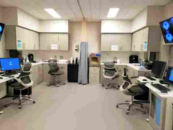
This set-up allows for simultaneous scanning procedures such as Hyperscanning – a neuroimaging technique that measures brain activity of two participants at the same time (with the two scanners are operating synchronously), allowing researchers to study how their brains interact during social interactions.

The stimulus presentation system available in both scanners provides projection of visual stimuli utilizing a high-resolution, long-life triple LED light source with a custom-designed, and mounted long-throw lens. Stimuli are projected onto a Velcro-mounted screen inside the scanner bore. Delivery of auditory stimuli occurs through stereophonic (dual-channel), air-coupled piezoelectric transducers surrounded by disposable bud-style canal tips that include passive noise reduction (28 dB attenuation). Each control room console unit includes amplifiers (with equalization), audio level with peak-detection limiters, and a multi-port video and audio input switch and multi-output VGA coupler (mirrors screen projection to a monitor in the scanner control room).
Participant responses are captured with non-magnetic, non-electronic response switches with up to five channels for multi-response paradigms, and a sixth channel processes triggering of the stimulus paradigm by the scanner once dummy scans are complete. Response switches and stereophonic earphones utilize optical fiber cables to transmit signal to the scanner control room. There are two MR compatible state-of the-art Eyelink 1000+ eyetrackers (SR Research) equipped with 2000Hz high-speed fiber-optic cameras available.
Our centralized control room was designed to accommodate information exchange between personnel, especially during sequence and protocol development as well as maintenance procedures.
Workroom at the Sammons BrainHealth Imaging Center, a Dallas-area location for advanced fMRI research including hyperscanning.

BIOPAC Modules

Our imaging center is also equipped with 2 BIOPAC MP160 data acquisition and analysis systems (Biopac Systems, Inc.) fully integrated with the Siemens MR scanners. The BIOPAC system with AcqKnowledge software + Basic Scripting is an Ethernet-ready modular data acquisition and analysis unit can record multiple (up to 16) channels with differing sample rates at speeds up to 400 kHz. The AcqKnowledge Acquisition & Analysis Software is integrated with the BIOPAC modules, allowing users to view, measure, transform, analyze, and report data.

Available to perform the following measurements:

  • electrical activity generated by the heart (normal ECG output and R-wave detection);
  • skin conductance level (SCL) and skin conductance response (SCR);
  • respiration (thoracic or abdominal);
  • adult human pulse oximetry;
  • SpO2 CO2 content in ambient air, expired breath, and in breathing gas;
  • heart rate (HR) data
  • relative central arterial pressure (providing noninvasive “Beat-by-Beat” Blood Pressure)

Additional Equipment