Overview
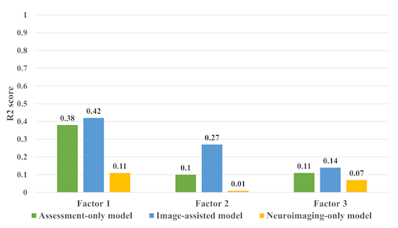
Read full research articleCan we use brain imaging data to better predict future brain health outcomes? The motivation is straightforward — the neural patterns and interactions identified through neuroimaging serve as early indicators that precede observable behaviors or psychological states — meaning a brain scan today could contain clues about cognition tomorrow. Cracking this code opens the door to earlier, smarter and more personalized interventions.This research collaboration developed and compared three different prediction approaches: 1) assessment-only, using support vector regression; 2) brain imaging alone via a random forest model; and 3) as their novel contribution, an "image-assisted" method that combined resting-state fMRI brain scans with behavioral assessments. This third approach used a sophisticated AI architecture called a partially conditional variational autoencoder (PCVAE), which essentially learns the relationship between brain connectivity and cognitive performance during training, then applies that learned knowledge to make future predictions using only the simpler assessment data — no repeat brain scan needed. The study included adult participants across two visits, allowing the team to assess both point-in-time predictions and how brain health changed over time. Results were promising: the image-assisted method outperformed both the assessment-only and neuroimaging-only approaches by effectively integrating neuroimaging data with assessment factors, suggesting that a one-time brain scan during training can meaningfully improve the accuracy of future predictions made without any imaging at all. This study underscores the potential of neuroimaging-informed predictive modeling to advance our understanding of the complex relationships between cognitive performance and neural connectivity. Looking ahead, key questions remain: How well do these models generalize across diverse populations, age groups, and clinical conditions? Can the approach scale to real-world clinical settings where MRI access is limited? And how many brain health "visits" are needed to build a reliably personalized model? Answering these will be essential before this kind of AI-assisted brain health forecasting becomes a routine tool in medicine.Below: Figure 5. The variation in R2 scores across factors 1, 2, and 3 between the assessment-only model, image-assisted model, and neuroimaging-only model for Visit 2 factor prediction. Higher R2 values are indicative of better performance. These findings imply that the image-assisted model has assimilated latent information from neuroimaging data during training, potentially enhancing its predictive capabilities when solely relying on assessment data in future predictions.