Read full research article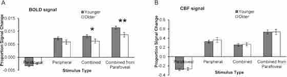
Overview
Researchers compared the blood flow in the brain to the metabolic rate of oxygen consumption in the brain using MRI scans. The rate of blood flow changes in comparison to the rate of oxygen consumption by cells in the brain as we age, which can cause cognitive performance to slow. This indicated that cognitive performance may depend on how efficiently neurons operate in the brain. When neurons are less efficient, they require greater oxygen levels to function correctly and more blood to bring that oxygen to the brain. Age-related changes in neuronal oxygen consumption and blood flow to the brain can explain the differences in blood-oxygen-level-dependent activation that lead to cognitive slowing.Figure 3. CBF and BOLD responses to visual stimulation. (A) BOLD percent signal change (mean ± SEM) for younger and older groups based on visual stimulation shown by stimulus condition. Significant differences between groups are indicated by asterisks: *P < 0.04, **P < 0.004. (B) CBF percent signal change (mean ± SEM) for younger and older groups based on visual stimulation shown by stimulus condition. (Comparison of the combined condition to parafoveal stimulation, shown in the rightmost bars of Fig. 3A,B, is aggregated from the parafoveal and combined stimulus conditions for both BOLD and CBF—that is, the effects are additive. Although this is not an independent measurement, it is shown here for illustrative purposes).