Facebook pixel
Go to home page

A Neural-Vascular Complex of Age-Related Changes in the Human Brain: Anatomy, Physiology, and Implications for Neurocognitive Aging

Senior man reading with a magnifying glass

Neuroscience & Biobehavioral Reviews

Dema Abdelkarim, Yuguang Zhao, Monroe P. Turner, Dinesh K. Sivakolundu, Hanzhang Lu and Bart Rypma

Read full research article
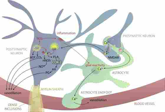
Overview

Functional magnetic resonance imaging (fMRI) plays a decisive role in testing hypotheses regarding the brain basis of age-differences in cognition and related mechanisms of neural-vascular coupling that affect cerebral blood flow. Researchers in this study reviewed current literature and controversies regarding the mechanisms of neural-vascular coupling and age-related changes. They concluded that nearly every component in this complex process is affected by aging, leading to pervasive age-related cognitive changes, including those affecting blood-oxygen level dependent signal (BOLD). This investigation generated novel questions about the roles of cellular networks in this process. Findings confirm how age-related changes in neural-vascular coupling can help elucidate causes of cognitive decline in older adults and reveal limitations of current scientific models.
Figure. 7. Older neurons. In aging, Ca2+ regulation by intracellular stores (e.g., the ER and mitochondria) is disrupted, and intracellular Ca2+ is higher as a result. Neuronal mitochondria produce more ROS with less regulation, causing damaging inflammation, and triggering glial reactivity. Myelin sheaths are thicker, with more layers, smaller internodes, and dense inclusions between lamellae. Abbreviations: AA, arachidonic acid; ATP, adenosine triphosphate; Ca2+, calcium; CO2, carbon dioxide; COX-2, cyclooxygenase-2; ER, endoplasmic reticulum; NMDAR, N-methyl-D-aspartate receptor; nNOS, neuronal nitric oxide synthase; PG, prostaglandin; PLA2, phospholipase A2; ROS, reactive oxygen species.

Figure. 7. Older neurons. In aging, Ca2+ regulation by intracellular stores (e.g., the ER and mitochondria) is disrupted, and intracellular Ca2+ is higher as a result. Neuronal mitochondria produce more ROS with less regulation, causing damaging inflammation, and triggering glial reactivity. Myelin sheaths are thicker, with more layers, smaller internodes, and dense inclusions between lamellae. Abbreviations: AA, arachidonic acid; ATP, adenosine triphosphate; Ca2+, calcium; CO2, carbon dioxide; COX-2, cyclooxygenase-2; ER, endoplasmic reticulum; NMDAR, N-methyl-D-aspartate receptor; nNOS, neuronal nitric oxide synthase; PG, prostaglandin; PLA2, phospholipase A2; ROS, reactive oxygen species.

Share this article


Bart Rypma, PhD

Principal Investigator Professor, Behavioral and Brain Sciences at UT Dallas Meadows Foundation Endowed Chair in Behavioral and Brain Sciences Director, Sammons BrainHealth Imaging Center


RELATED INFORMATION

Sammons BrainHealth Imaging Center

Located at Center for BrainHealth, the Sammons BrainHealth Imaging Center is a one-of-a-kind facility completely focused on human brain imaging to measure changes in brain health and function. The design and field strength are optimized for functional MRI (fMRI), providing crisp anatomical detail and a signal-to-noise ratio that reduces distortion to facilitate the acquisition and interpretation of data.

Altered Linear Coupling Between Stimulus-Evoked Blood Flow and Oxygen Metabolism in the Aging Human Brain

Human aging affects the structural integrity of neural-vascular coupling (NVC), which plays a critical role in healthy neural function by delivering oxygen and nutrients to metabolically active neurons.