Facebook pixel
Go to home page

BOLD Hemodynamic Response Function Changes Significantly with Healthy Aging

Happy elderly senior couple jogging running or walking outside on a wooded trail. Older.

NeuroImage

Kathryn L. West, Mark Zuppichini, Monroe P. Turner, Dinesh K. Sivakolundu, Yuguang Zhao, Dema Abdelkarim, Jeffrey S. Spence and Bart Rypma

Read full research article
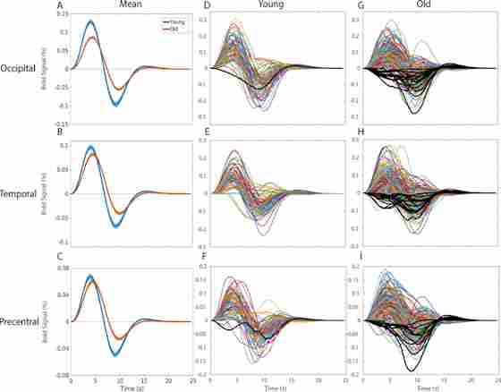
Overview

Hemodynamic response function (HRF) characterizes the blood-oxygen-level-dependent (BOLD) signal of the brain over time through functional magnetic resonance imaging (fMRI). HRF can change over time as we age, and this is an important measurement in studies of the aging brain and its function. This study sought to use a large sample size to assess the HRF changes in healthy aging participants. The HRF was calculated in the visual, auditory, and motor cortices and there was found to be a significant difference for all three between older and younger participants. These neurovascular changes due to ages are relatively new findings, and further studies will continue to explore the implications of such discovery.
Figure 2. Hemodynamic Response Functions across (blue) young and (orange) healthy older adults in A) the occipital region of interest in response to a checkerboard stimulus for 34 ms, B) the temporal region of interest in response to 300 ms binaural tone at 1200 Hz, and C) the LH precentral region of interest due to right-index finger button-press response with the (solid line).

Figure 2. Hemodynamic Response Functions across (blue) young and (orange) healthy older adults in A) the occipital region of interest in response to a checkerboard stimulus for 34 ms, B) the temporal region of interest in response to 300 ms binaural tone at 1200 Hz, and C) the LH precentral region of interest due to right-index finger button-press response with the (solid line).

Share this article


Jeffrey S. Spence, PhD

Director of Biostatistics

Bart Rypma, PhD

Principal Investigator Professor, Behavioral and Brain Sciences at UT Dallas Meadows Foundation Endowed Chair in Behavioral and Brain Sciences Director, Sammons BrainHealth Imaging Center


Related Information

Sammons BrainHealth Imaging Center

Located at Center for BrainHealth, the Sammons BrainHealth Imaging Center is a one-of-a-kind facility completely focused on human brain imaging to measure changes in brain health and function. The design and field strength are optimized for functional MRI (fMRI), providing crisp anatomical detail and a signal-to-noise ratio that reduces distortion to facilitate the acquisition and interpretation of data.

Is your brain as fit as you are?

Three decades of science have shown that you can train your brain to work better, faster and last longer. When you join The BrainHealth Project, you take your BrainHealth Index (BHI), get your personalized training plan, and explore your brain’s upward potential. It’s all online, and you can access it from anywhere.