A collaboration between The University of Texas at Dallas' Center for BrainHealth® and UT Southwestern Medical Center has resulted in a new way to look at the brains of patients with multiple sclerosis (MS) which could greatly enhance doctor's ability to select the best therapy for each person.
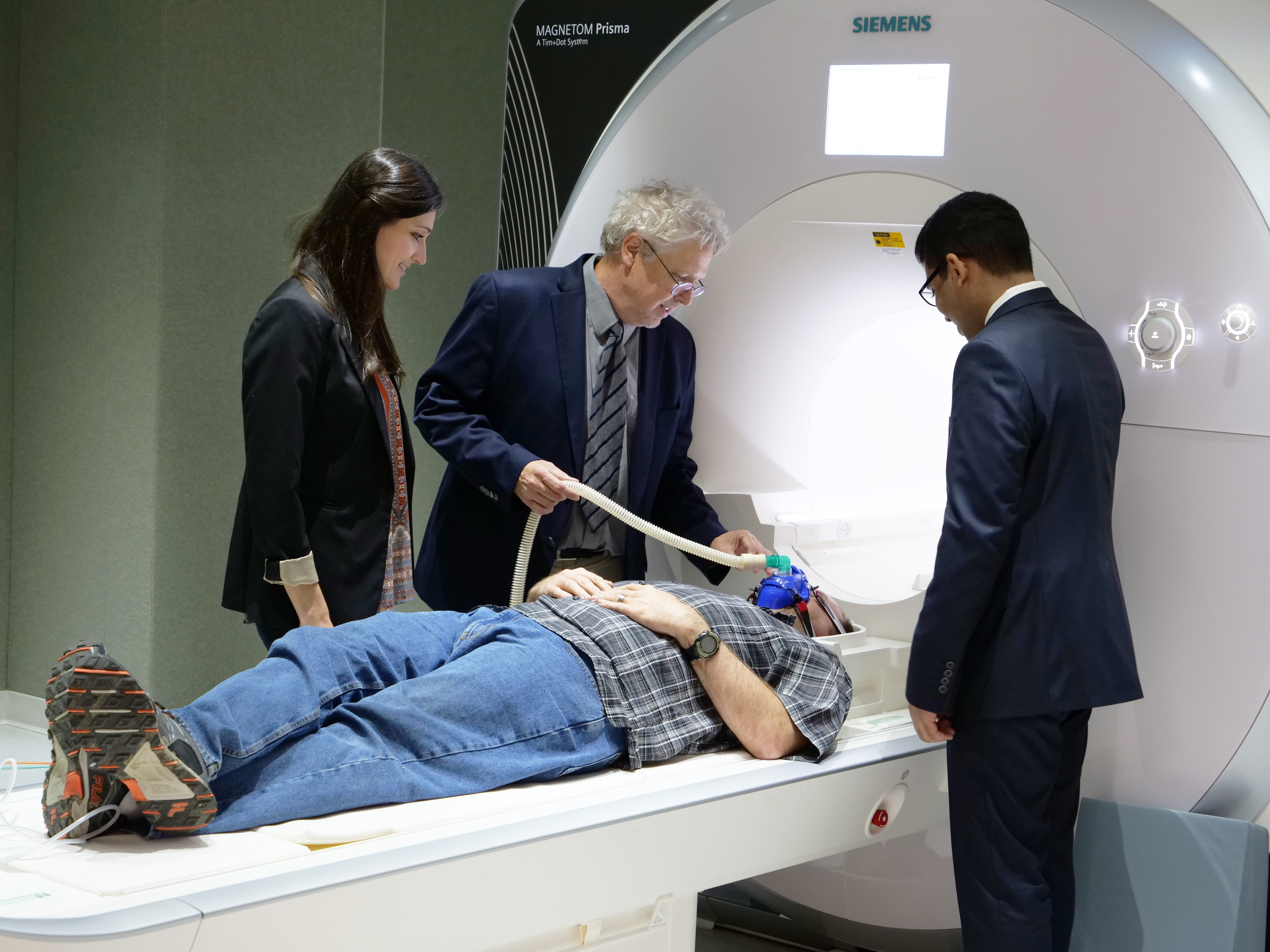
In a study published in the Journal of Neuroimaging, researchers were able to examine brain lesions using a patent-pending technique employing 3 Tesla MRI, allowing doctors to see the lesions in 3D instead of 2D. Previously, doctors were only able to look at lesions in 2D, forcing doctors to make educated guesses on the status of each lesion.
Having a 3 dimensional view of the lesions allows doctors to see the harm done to the tissue surrounding the lesions and allows doctors to give a better estimate on how long the lesion has existed.
“”
"It is exciting because prior to this development, doctors have had to rely on trial and error therapy. So they just look at the lesion and make their best guess about it," said Dr. Bart Rypma, a professor of psychology at UT Dallas. "So with our technology we can distinguish a newer lesion that have more recently occurred and have the potential to heal themselves and older lesions that tend to be in a more static state and are less capable of healing."
Read full story on KRLD