
Read full research article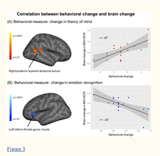 These findings are preliminary, and further research is needed to determine the extent of the VR-SCT program’s potential. However, findings from this pilot study provide valuable insights on ways the neuroplasticity of individuals with ASD can be harnessed.
Overview
Research prior to this pilot study has relied primarily on behavioral observations, with limited information on the specific neural mechanisms at play during treatments for individuals with autism spectrum disorder. A Phase I trial of this pilot study targeted this void and investigated the impact of an evidence-based social training program on the brain. The program utilized, Virtual Reality-Social Cognition Training (VR-SCT), was developed at the Center for BrainHealth® at UT Dallas and has produced promising results in previous studies. What made this study’s approach novel was the focus on how the participants’ brains changed after VR-SCT. Using a well-validated social perception fMRI measure on young adults with ASD, researchers identified three main brain changes. First, an area of the brain that serves as a hub for socio-cognitive processing (right posterior superior temporal sulcus), showed increased activation in individuals that showed improvements in their theory-of-mind measure. In other words, for the participants that showed improvements on measurements related to discerning the intentions of others, researchers saw greater levels of activation in the socio-cognitive processing brain region. Second, an area associated with socio-emotional processing tracked gains in emotion recognition. Lastly, a brain region utilized for visual attention showed significantly decreased activation to social vs. nonsocial stimuli for all participants, where heightened attention to nonsocial contingencies has been considered a disabling aspect of ASD.Figure 3 - The portion of the brain highlighted in red, the right posterior superior temporal sulcus (pSTS), is an area associated with the integration of visual, auditory and somatosensory cues of other’s behaviors. This is an important piece of social interaction, as it allows us to process incoming information and make predictions about what we think someone will do next. Decreased activation in the portion of the brain highlighted in blue, the left inferior frontal gyrus, was shown to be associated with gains in participants’ socio-emotional processing of identifying emotions.

